
.png)


The vendor-neutral field guide to how medical images are created, stored, moved, displayed, and shared across the modern health enterprise. Written for everyone who touches imaging, not just radiologists.
15 page PDF 5 parts 14 glossary terms 20 minute read
The framework
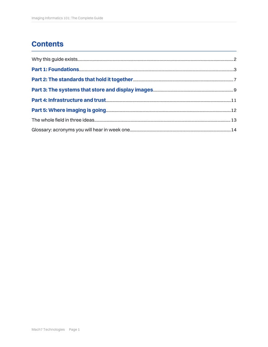
- 01 Foundations Free to read below
- 02 The standards that hold it together In the download
- 03 The systems that store and display images In the download
- 04 Infrastructure and trust In the download
- 05 Where imaging is going In the download
- The whole field in three ideas In the download
- Glossary: acronyms you will hear in week one In the download
Why this guide exists
Medical imaging is the largest data type in healthcare, and one of the least understood outside the reading room. A single hospital produces studies from dozens of devices, stores them for decades, moves them between facilities, and puts them in front of clinicians who are rarely radiologists.
Enterprise imaging data can account for up to roughly 90% of a health system's total storage consumption, and hospitals generate on the order of 50 petabytes of data per year. HealthTech Magazine, 2024
This guide is the map. It explains imaging informatics from first principles, in language a new engineer, a clinical champion, an IT administrator, or a curious executive can follow. It is vendor neutral by design. The goal is understanding, not a sales pitch.
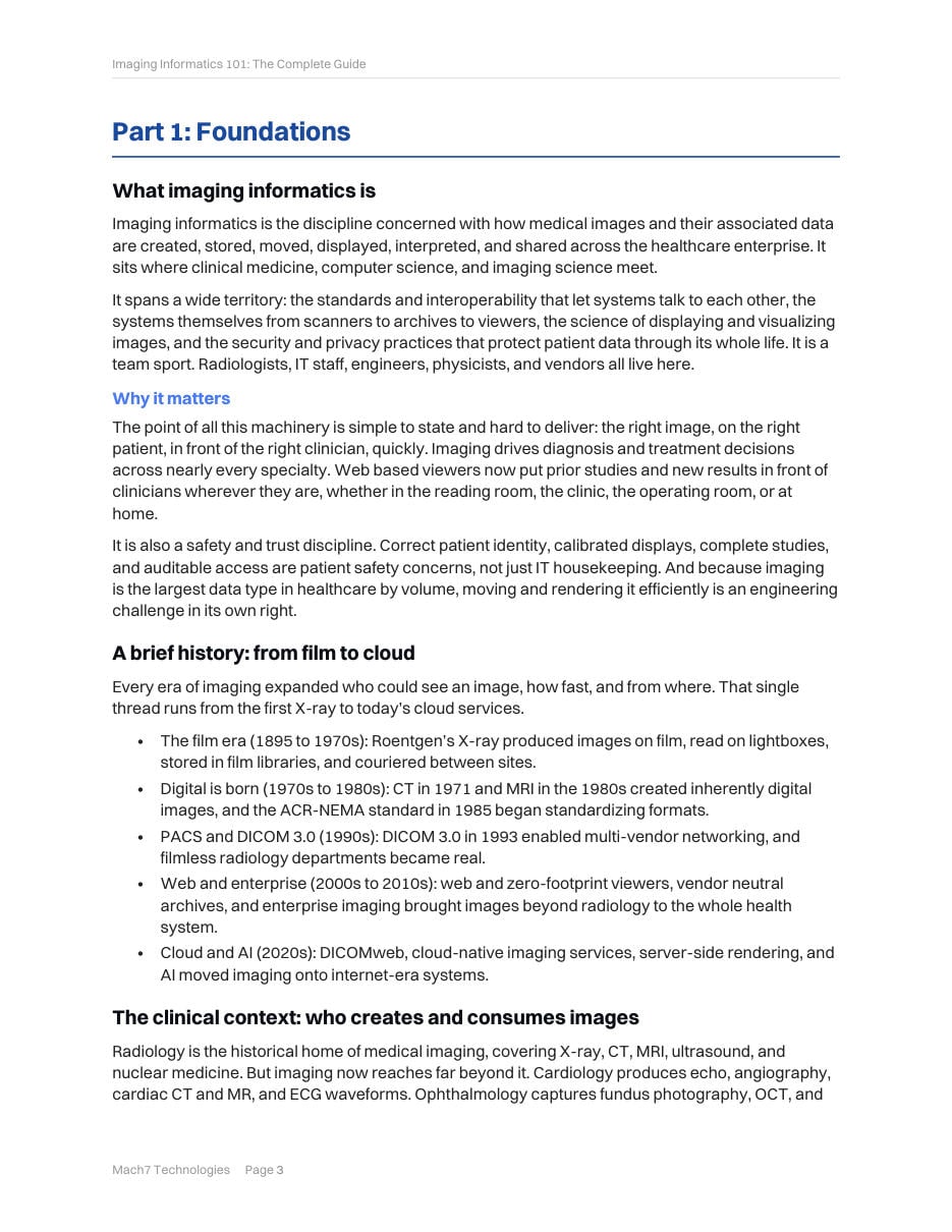
Part 1: Foundations
What imaging informatics is
Imaging informatics is the discipline concerned with how medical images and their associated data are created, stored, moved, displayed, interpreted, and shared across the healthcare enterprise. It sits where clinical medicine, computer science, and imaging science meet.
It spans a wide territory: the standards and interoperability that let systems talk to each other, the systems themselves from scanners to archives to viewers, the science of displaying and visualizing images, and the security and privacy practices that protect patient data through its whole life. It is a team sport. Radiologists, IT staff, engineers, physicists, and vendors all live here.
Why it matters
The point of all this machinery is simple to state and hard to deliver: the right image, on the right patient, in front of the right clinician, quickly. Imaging drives diagnosis and treatment decisions across nearly every specialty.
Just as important are the consumers: referring physicians, surgeons, oncologists, emergency teams, tumor boards, and increasingly patients themselves through portals. The key insight for anyone building or running imaging systems is that most viewer users are not radiologists.
Imaging modalities
Imaging devices fall into five broad families. The two-letter codes in parentheses are DICOM modality codes, and you will see them everywhere.
- Projection radiography (CR, DX, MG): X-ray passes through the body onto a detector.
- Cross-sectional (CT, MR): the body reconstructed as slices or volumes.
- Real-time and dynamic (US, XA, RF): live, moving images during an exam or procedure.
- Functional and molecular (PET, SPECT, NM): physiology and metabolism, not just anatomy.
- Visible light and specialty (OCT, fundus, whole-slide, endoscopy): cameras and optical scanners.
| Modality | Ionizing | Form | Typical study size | What that means for software |
|---|---|---|---|---|
| X-ray (CR, DX) | Yes | 2D projection | 10 to 50 MB | Small and fast, but enormous volume |
| Mammography (MG, DBT) | Yes | 2D plus pseudo-3D | 50 MB to 3 GB | High-resolution display, priors side by side |
| CT | Yes | 3D slice stack | Hundreds of MB | Fast scroll, multi-planar and 3D, many slices |
| MRI | No | 3D, multi-sequence | Hundreds of MB | Many series, so hanging protocols matter |
| Ultrasound and echo | No | 2D plus cine loops | Tens of MB | Smooth video playback |
| PET/CT | Yes | Functional plus anatomic | Hundreds of MB | Fusion overlay of two datasets |
| OCT (ophthalmic) | No | 3D B-scan volume | Tens to hundreds of MB | Frame-to-fundus correlation |
| Whole-slide (SM) | No | Gigapixel 2D pyramid | Several GB | Tiled, progressive streaming |
Sizes are approximate and highly variable. The point is the range: roughly four orders of magnitude between a chest X-ray and a whole-slide image.
Look inside
The page before the glossary compresses the whole field into three sentences. They are the reason the other fourteen pages stick.